Cardiology Case #5
Primary Author: Dr Alastair Robertson; Co-Authors: Dr Hywel James and David Law
Background:
72 year-old male presents with increasing breathlessness over the last 2-3 days which was preceded by an episode of central chest heaviness which occurred whilst doing some gardening.
He has a background of cardiac sarcoid (on immunosuppression) and has a permanent pacemaker, inserted for complete heart block.
He is pain free with normal observations. RR 18, Sats 96%, BP 105/69
Initial ECG
-
Paced rhythm with a rate of 85. There a dual pacing spikes indicating both atrial and ventricular pacing.
The QRS is relatively narrow, around 100-120ms suggesting pacing of the conduction system.
ST segments are challenging to analyse in paced rhythms. There is ST elevation leadII, V2-V6 but no clear reciprocal changes.
This is a concerning ECG in the setting of dyspnoea and chest pain.
Although there was no further chest pain, there were evolving ECG changes with dynamic ST segments.
Initial hsTroponin was 2,500.
-
Differentials to consider include:
Ischaemia - evolving STEMI
LV aneurysm
Cardiomyopathy due to sarcoid
Myopericarditis
Stress Induced [Takotsubo] cardiomyopathy
Aortic dissection
PE
Cardiac POCUS
The first video shows a Parasternal Long Axis (PLAX) View.
The second video is a Parasternal Short-axis (PSAX) at the base of the LV (just distal to the mitral valve).
The final video shows a PSAX at the LV mid/apex region.
What do you think of the two short axis clips?
-
PLAX - Basal LV is functioning well, aortic and mitral valves appear normal, RV and aortic root size look normal, and there is a very small pericardial effusion.
Looking closer at the LV, it appears hyperdynamic at the base, but the apical portion shows dilatation with hypokinesis of both the anterior septum (LAD territory) and the inferior wall (RCA/LCx territory).
PSAX at the base (first clip) shows very dynamic, symmetrical contractility.
PSAX towards the apex (second clip) shows marked global hypokinesis of the entire apex.
These initial views show apical ballooning and hypokinesis, with a preserved and hyperdynamic LV base. This could be consistent with MI affecting the apex (due to LAD occlusion), but could also be suggestive of stress induced [Takotsubo] cardiomyopathy.
KEY at this point are obtain apical views to assess if the LV impairment maps a particular vascular territory.
Could this be Cardiac Sarcoid?
Cardiac sarcoid can cause a cardiomyopathy but was thought less likely to be the cause of his symptoms. Typical echo findings of sarcoid are LV dilation, septal thinning, and hypokinesia of the left ventricle that usually localise to the proximal anterior septum (or lateral free wall). In the long-axis we can see the proximal anterior septum is hyper-kinetic.
Apical Views
Here are the Apical 4-chamber (top), Apical 2-chamber (middle) and Apical long-axis (bottom).
The Apical long-axis view (sometimes called ‘Apical 3-chamber’ is the same as the PLAX but with improved visualisation of the apex.
POCUS Pearls:
All three of the clips above show ‘ballooning’ or dilatation of the mid- and apical- segments of the LV whilst the base is hyperdynamic with symmetrical contraction. This pattern is global and did not fit a particular vascular territory so consideration was given to Takotsubo cardiomyopathy rather than acute ischaemia.
It is important to note however, that Takotsubo is a diagnosis that can only be made after angiogram. You must assume its an acute coronary event [e.g LAD occlusion] until proven otherwise!
Basic POCUS:
Again, resort back to a good PLAX - can tell you a lot about the cardiac function.
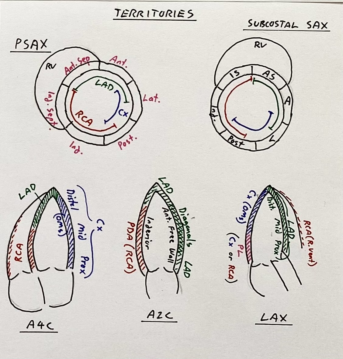
Space repetition for the vascular territories (see below). Here the mid-LV and apical LV is hypokinetic in all of the views. Look for ballooning of the apex.
Let’s discuss the views and the territories….
In the apical-4C the lateral wall (on right, LCx territory) as well as the distal septum (on left, LAD) are impaired.
In the apical-2C the anterior free wall (on right, diagonals [from LAD]) as well as the posterior wall (on left, PDA/RCA) are impaired.
In the apical long-axis the anterior septum (on right, LAD) as well as the inferior wall (on left, LCx via OMs) is impaired.
Compare this global impairment to the territorial LV impairment seen in Case 1 (large LAD STEMI).
Vascular territories on the key Apical, and Short-axis views.
Intermediate POCUS:
Takotsubo is a diagnosis of exclusion, confirmed after angiogram but in cases such as this echo can be useful to assess for complications.
Apical hypo- or akinesis is a significant risk for the development of LV thrombus. Carefully assess the apex (top video) for evidence of LV thrombus by fanning through from wall to wall (see Case 3 - large LV thrombus)
Another complication of Takotsubo can be LV outflow tract obstruction, precipitated by the hyperdynamic LV base obstructing the outflow as it contracts during systole. Carefully assess for the mitral valve being pulled anteriorly to obliterate the outflow tract (in PLAX).
POCUS progression:
Quantitative methods of assessing for LVOTO are high-peaking “dagger-like” doppler signal through the LVOT or elevated gradients across the LVOT.
Case Conclusion
Serial troponins were downtrending and the patient remained pain free. Further questioning revealed some significant recent social stressors prior to his symptoms onset.
Angiogram showed double-vessel disease which was stented, but report comments that angiographic findings were suggestive of Takotsubo cardiomyopathy.
There was no clear consensus from our Cardiology team as to whether this was Takotsubo or coronary artery occlusion, showing how difficult a diagnosis it can be where they can potentially coexist.
Extra Tips:
Always consider LV Outflow Tract Obstruction in shocked patients with pathologies such as:
HOCM or Hypertensive cardiomyopathy
Takotsubo cardiomyopathy
Apical hypokinesis from LAD myocardial infarction
Cor Pulmonale
Significant hypovolaemia
Conventional therapies with inotropes such as Adrenaline may make them worse. What these patients often need is (cautious) fluids to increase preload and rate control to allow time for adequate LV filling.
Spotlight on…..Cardiac Sarcoid
Sarcoid is a granulomatous disorder than can occur in any organ. In the heart it often causes AV blocks (hence this patient having a PPM inserted), valvular pathology, cardiomyopathy or CCF. Echo classically shows LV dilatation, septal thinning and segmental impairment, usually anterior septum or lateral free wall. Diagnosis may require scintigraphy, PET, or MRI.


